An interdisciplinary study
Mummies bear witness to a millennia-old past. Analysis of the human remains concealed beneath their wrappings allows us to rediscover fragments of past lives, ancient illnesses, and the embalming techniques that were used to prepare the bodies of the deceased for eternal life.
Today, scholars have cutting-edge technologies at their disposal that allow for non-invasive analyses which include imaging investigations such as computed axial tomography (CT), which allows mummies to be virtually 'unwrap' as well as facilitating palaeoanthropological studies.


The mummy with the painted shroud underwent a CT scan thanks to a collaboration with the Radiology Department of the IRCCS Azienda Ospedaliero-Universitaria di Bologna. The CT scans were performed when the ward was closed to the public. The mummy was transported inside a special sealed container called a Conservation Soft Box which was designed and conceived by the Eurac Research Institute for Mummy Studies. The Conservation Soft Box made it possible to protect the mummy, control the correct conservation parameters and avoid contaminating the hospital environment.


The mummy in the Conservation Soft Box
© PAOLO BONDIELLIThe CT analyses
The mummy contains the remains of a woman, about 153 centimeters tall, who may have been 35-45 years old at the time of death. Analysis did not reveal a single cause of death. The woman was afflicted with abscesses that resulted in the loss of some teeth during her lifetime. She also had degenerative diseases, such as osteoarthritis of the spine and knee joints.
The body of the woman is in a supine position, with her arms stretched out along her sides and straight legs.
The abundant folds of the skin and the residue of fatty tissue on the hips, buttocks, and thighs suggest the individual may have been overweight.


Some of the details of the face painted on the shroud appear opaque or radiodense in these 3D reconstruction images. This is because these particular facial features as well as many other of the shroud’s details, were painted with iron-based pigments that inhibit the passage of CT X-rays.
Embalming techniques
Virtual CT scanning of the mummy revealed that the body is in a supine position with the arms stretched along its sides and the legs straight. During the embalming process, the brain was almost completely removed through the left nostril. The internal organs were extracted through a vertical incision in the abdomen, which was then only partially stuffed with resin-soaked bandages. The body was finally covered with a thick resin cast and then with a linen bandage.


The internal organs were extracted through a vertical incision in the abdomen, which was then only partially stuffed with resin-soaked bandages.
In preparation for bandaging, the limbs of the deceased were secured with knotted fabric bands at the wrists, under the pubis and below the knees. The body was then generously covered with a cast of resin and then overlayed with a linen cloth bandage.
Radiocarbon dating

Fragment for the radiocarbon dating
© Eurac Research - Marco SamadelliRadiocarbon dating (¹⁴C), carried out on a fragment of the painted shroud and another fragment of the inner bandage, attributes the piece to the Roman era (1st-2nd century AD). Stylistic comparisons with mummies and sarcophagi belonging to members of the Soter family, whose tomb was found in the Theban necropolis of El-Khokha (TT32), point to the same period.


Painted shroud of Sensaos, Roman period, reign of Trajan. Soter tomb, Thebes.
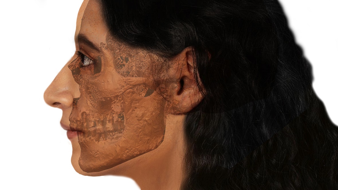
Craniofacial reconstruction
Craniofacial reconstructions restore the facial features of individuals who lived in the past as realistically as possible. To create them, an excellent knowledge of facial anatomy and an assessment of the mechanical interactions between the soft tissue and the underlying bone structure are indispensable. The virtual anthropologist considers the characteristics of facial bones by combining them with measurements of numerous standardized points on the skull to calculate facial volumes. The data is then combined with existing information on the average tissue thickness of human faces, which differs between populations.
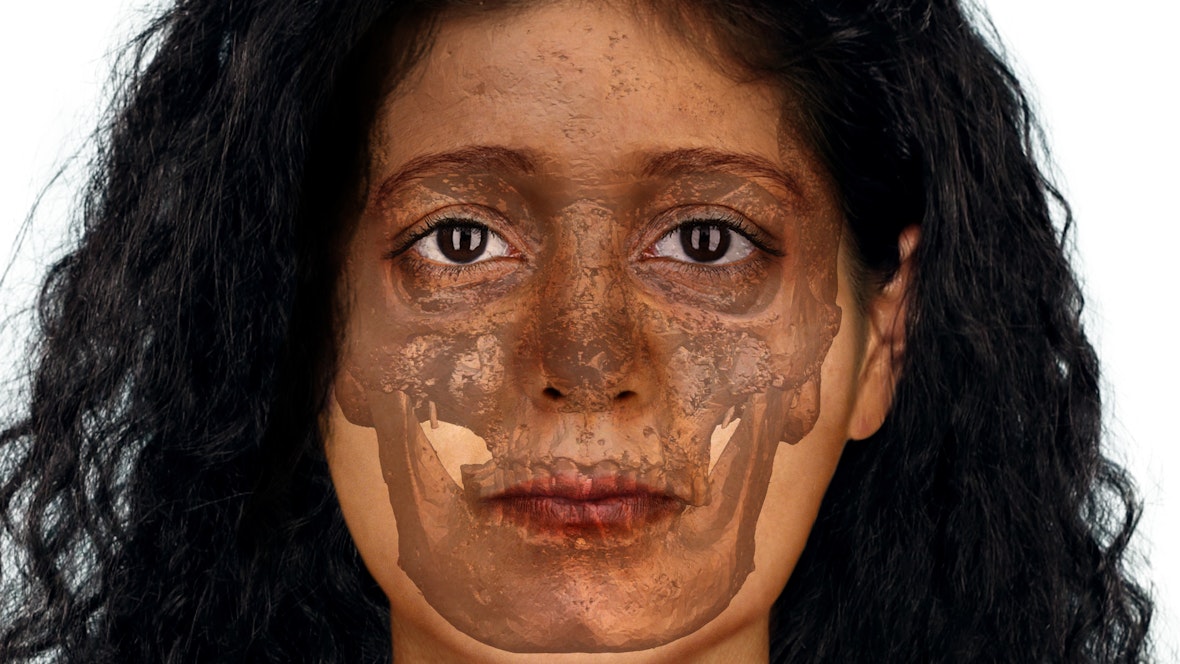
Shift the vertical line to show the facial reconstruction
© Viviana Conti, Lucrezia Rodella
To reconstruct the face of the mummy with the painted shroud, a digital model of the skull which had been acquired by CT scan, was created. Using special 3D modelling software, frontal and left lateral images of the skull were obtained and the facial features were estimated. The final result was produced by combining images of individuals with similar facial features to those of the deceased. Genetic results, obtained by the Institute for Mummy Studies, of other coeval Egyptian individuals were considered for the choice of eye, skin and hair colour for the mummy with the painted shroud.
Shift the vertical line to show the facial reconstruction
© Viviana Conti, Lucrezia Rodella
