magazine_ Article
The beauty of structure
A neuroscientist explains fascinating advances in microscopy.
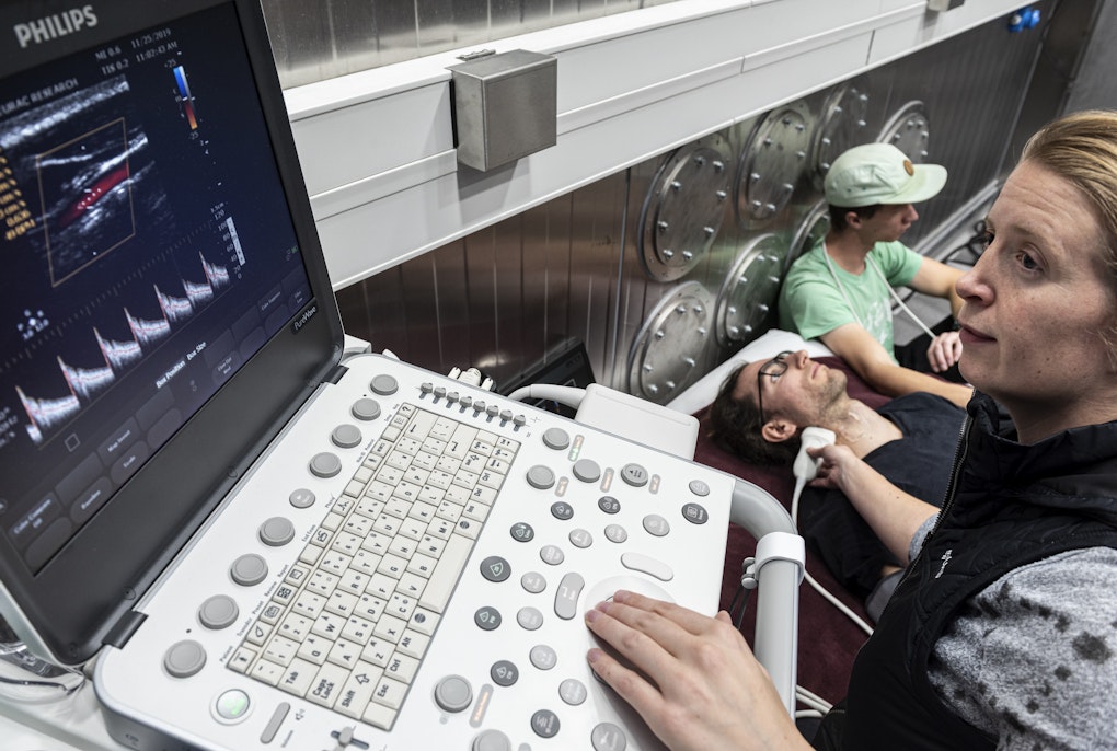
Using sophisticated confocal microscopy systems, Alexandros A. Lavdas provides cellular insights that are fundamental to a better understanding of processes and mechanisms.
Photo: Ivo Corra' | Eurac Research
Neuroscientist Alexandros A. Lavdas is not only an expert in imaging he’s also a happy man: his insights into the inner life of cells not only satisfy his drive for knowledge as a researcher, but also give him great aesthetic pleasure.
When neuroscientist Alexandros Lavdas describes images of the cerebral cortex – “there's this six-layered structure, and each layer is made up of different cells, and the whole thing looks beautiful and very intricate and artistic” – he sounds as enthused as he does describing the close-up photos he takes of leaves or fruits: “With the right lens, the most fantastic details become visible!”
Lavdas is an expert in imaging in biomedical research and a passionate amateur photographer, and both are related to his pronounced “proclivity for images”. As a student this passion made him stand out: “Most of my student friends were crazy about molecular biology,” says the scientist, who grew up in Athens as the son of two classical musicians. “I didn't have the slightest interest in it. I know of course, it's terribly exciting and absolutely essential – it's just not for me.” His interest was in morphology, anything to do with shapes. And this affinity, combined with his enthusiasm for photography, enabled him to “understand concepts of imaging that most bio-scientists were not really all that interested in.” Although he grew up in an artistic home “needing art as much as oxygen”, he knew from a young age that he would follow the path of science. For a while he thought about physics; when he finally decided on biology, it was with the clear goal of becoming a neuroscientist.
At University College London he carried out research on cortical development, and later, at the Pasteur Institute in Athens, on injury and repair of the central nervous system – “but even by then, I was already the unofficial go-to-person for imaging; everyone came to me for help and advice because I had the most experience with it.”
If you want to publish results, you have to quantify. Image software can not only quantify results, but it can make them clearly visible.
Alexandros Lavdas
Officially however, he did become responsible for imaging at the Eurac Research Institute for Biomedicine, where he set up the facility from scratch in 2014. With his knowledge and sophisticated confocal microscope systems, he facilitates cell insights that are fundamental for a better understanding of processes and mechanisms, both with regard to the emergence and development of diseases and the effect of drugs and while doing so, derives aesthetic not only pleasure but knowledge from this: “Oh yes, I really enjoy the images.”
In the analysis, of course, the numbers come into play. “If you want to publish results, you have to quantify. But the image software, apart from the quantification, can make results very clearly visible. I can color code and specify making values above a certain level, green for example. And suddenly you see green popping up in the image and you know that you have the values you're looking for.”
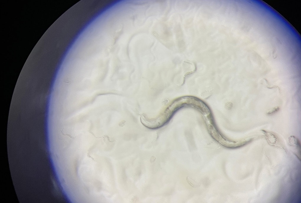
Lavdas often examines mitochondria, the small organelles that supply the cell with energy, because their condition tells a lot about cell health. Fragmentation is a bad sign. “I can tell the software to give the shorter fragments a cooler color. And then looking at two cells if one is bluer and the other redder, I can tell redder one is healthier.”


Lavdas also gives mitochondria as an example of how improved technology changes our view of reality. From the illustration in his university textbook, one had to get the impression that these organelles were scattered here and there in the cell, “because that's what they look like under the electron microscope.” For electron microscopy, however, samples undergo a procedure (“torture”, Lavdas says) that take them quite far from their natural state: they are fixed, dehydrated, and then embedded in plastic - “they go into the oven at 70 degrees, are exposed to osmium ... these cells go through hell!” He laughs. He can take videos of mitochondria in living cells today, “and you can see, they are everywhere! From the edge of the nucleus to the edge of the cell, a network of branching tubes that form and break and form again, there's a dynamic.” The beauty of the mitochondria inspires him, as does that of neural tissue.
If these possibilities had been described to life scientists a few decades ago, they would have seemed like science fiction to most of them.
Alexandros Lavdas
Lavdas has experienced something of a revolution in imaging since a cell biology professor at the University of Athens let him work alone at the electron microscope as a 21-year-old undergraduate (which made some people on the faculty feel a bit uncomfortable). “The revolution in the last few decades is about visualizing cells and samples in high enough resolution – not as high as electron microscopy, but high enough –in a state that is much more natural. “ Advances in many fields have contributed to this development, not least immensely increased computer power. Years ago, a whole computer storage drive would not have been enough to hold the data of an experiment like the ones the institute now does every day. In optics, there have also been decisive developments in confocal microscopy, “tomography for cells”, as Lavdas explains: “The sample is optically cut into layers, usually with lasers, which the computer then can reconstruct in 3D; after you mark different proteins with antibodies with different colors, you can turn the cell around and look at it from all sides. Like this, you get a wealth of information about the morphology and the contents of the cell.” If these possibilities had been described to bio-scientists a few decades ago, they would have “seemed like science fiction to most”, Lavdas says. “And all these things are getting better and better; the sensitivity of the detectors is increasing, so you need less intense lasers, which means less damage to the cell.” He has himself worked in a collaborative project between Eurac Research, the Politecnico di Milano, the University of Cambridge and the Bolzano-based company Micro Photon Devices to develop a novel system which uses “FLIM imaging”, a technique that helps us acquire biochemical information from the cells.
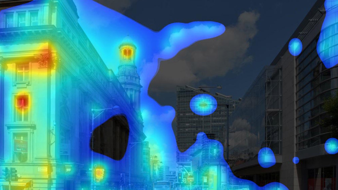
Heat map of first gaze probability (3-5 sec after image presentation) in an urban scene, as computed by artificial intelligence software trained with actual eye-tracking data. Warmer colors represent a higher probability to attract the first gaze, cooler colors a lower probability, while areas with no color are not likely to attract the gaze at all. Such heatmaps help us understand what morphological features attract our gaze in buildings or other urban or natural scenes.Photo: Eurac Research / Alexandros Lavdas Photo: Eurac Research / Alexandros Lavdas
But apart from imaging, Alexandros Lavdas is also occupied with another major topic: neuroaesthetics. More specifically, he is researching how our brain perceives shapes, which shapes we find beautiful, and for what reason. In a study with landscape ecologist Uta Schirpke two years ago, he showed that we are particularly attracted to forms with certain types of organized complexity, forms that have been proven to have a positive effect on our well-being. This applies to landscapes (the subject of his most recent paper with Schirpke and Erich Tasser) as well as to architecture – a field in which Lavdas is particularly interested since buildings are among his favorite photographic subjects. His most recent papers are about developing quantitative tools for measuring the appeal and effect of buildings on humans. Since last year, he has also been a member of the board of directors of the Human Architecture and Planning Institute, an organization in the US dedicated to understanding the human experience of the built environment. Alexandros Lavdas is quite literally dealing with the shape of things to come.